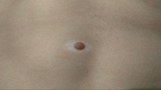
Precīzs halo nevusa cēlonis nav pilnībā izprasts, bet tas ir saistīts ar imūnsistēmas uzbrukumu pigmenta šūnām dzimumzīmē un ap to. Ģenētika, vitiligo, autoimūnas slimības un UV starojums var spēlēt lomu. Rezultāts ir pigmenta zudums ap dzimumzīmi, veidojot balto halo.
Pats par sevi halo nevus tiek uzskatīts par drošu un būtiski nepalielina melanomas risku. Galvenā problēma ir, ja dzimumzīme halo iekšpusē izskatās netipiska vai sāk ātri mainīties. Jebkuras ātras izmaiņas, jauni simptomi vai ļoti neregulāras pazīmes būtu jāpārbauda dermatologam.
Lielākajai daļai halo nevusu nav nepieciešama ārstēšana, un tos var vienkārši novērot laika gaitā. Ja dzimumzīme izskatās aizdomīga, tiek atkārtoti traumēta vai mainās satraucošā veidā, ieteicama ķirurģiska noņemšana ar histoloģiju. Destruktīvas metodes, piemēram, lāzers vai sasalšana, nav ideālas, jo tās iznīcina audus, kas nepieciešami pareizai pārbaudei.
Tu nevari pilnībā novērst halo nevusus, jo tos izraisa tava imūnsistēma un ģenētika, bet tu vari aizsargāt savu ādu kopumā. Ierobežo UV starojumu, izvairies no solārijiem un izmanto sauļošanās krēmu un apģērbu, lai mazinātu saules bojājumus. Centies izvairīties no hroniskas berzes vai traumas dzimumzīmēm un seko līdzi jebkurām, kas mainās.
Apmeklē dermatologu, ja halo nevus izskatās ļoti atšķirīgs no tavām citām dzimumzīmēm, ātri mainās vai sāk niezēt, sāpēt, asiņot vai veidot kreveles. Regulāras pārbaudes ik pēc 1–2 gadiem ir laba ideja, ja tev ir daudz dzimumzīmju, ādas vēža vēsture vai vairāki halo nevusi.
Tipisks, stabils halo nevus nav ārkārtas situācija un to var pārbaudīt ikdienas dermatoloģijas vizītē. Ja dzimumzīme halo iekšpusē ātri mainās, izskatās ļoti neregulāra vai sāk niezēt, sāpēt vai asiņot, tev vajadzētu noorganizēt neārkārtas, bet steidzamu tikšanos dažu nedēļu laikā.