What Is a Spitz Nevus?
A Spitz nevus is a distinctive type of melanocytic nevus composed of large, epithelioid and spindle-shaped melanocytes that was first described by Dr. Sophie Spitz in 1948. Originally termed juvenile melanoma because of its histologic resemblance to melanoma, the name was changed to Spitz nevus when it became clear that the vast majority of these lesions are benign despite their alarming microscopic appearance.
Spitz nevi account for approximately one percent of all melanocytic nevi in children and are most commonly diagnosed in patients under the age of 20, with a peak incidence in the first decade of life. However, they can occur at any age including in adults. Clinically, a classic Spitz nevus presents as a smooth, dome-shaped, pink or red papule or nodule — typically measuring five to ten millimeters in diameter — that appears suddenly and may grow relatively rapidly over weeks to months before stabilizing.
The characteristic pink or reddish color results from the prominent vascularity within the lesion. Some Spitz nevi are pigmented (brown to black), and these pigmented variants — sometimes called Reed nevi or pigmented spindle cell nevi — are more common in adults and on the lower extremities. The most common location is the face, particularly the cheeks, followed by the extremities.
Spitz nevi are typically solitary, though rare agminated (grouped) or disseminated variants exist. The clinical significance of Spitz nevi lies not in any inherent danger but in the diagnostic challenge they pose — their clinical and histologic overlap with melanoma makes definitive classification one of the most contentious areas in dermatopathology.

Why Spitz Nevi Mimic Melanoma
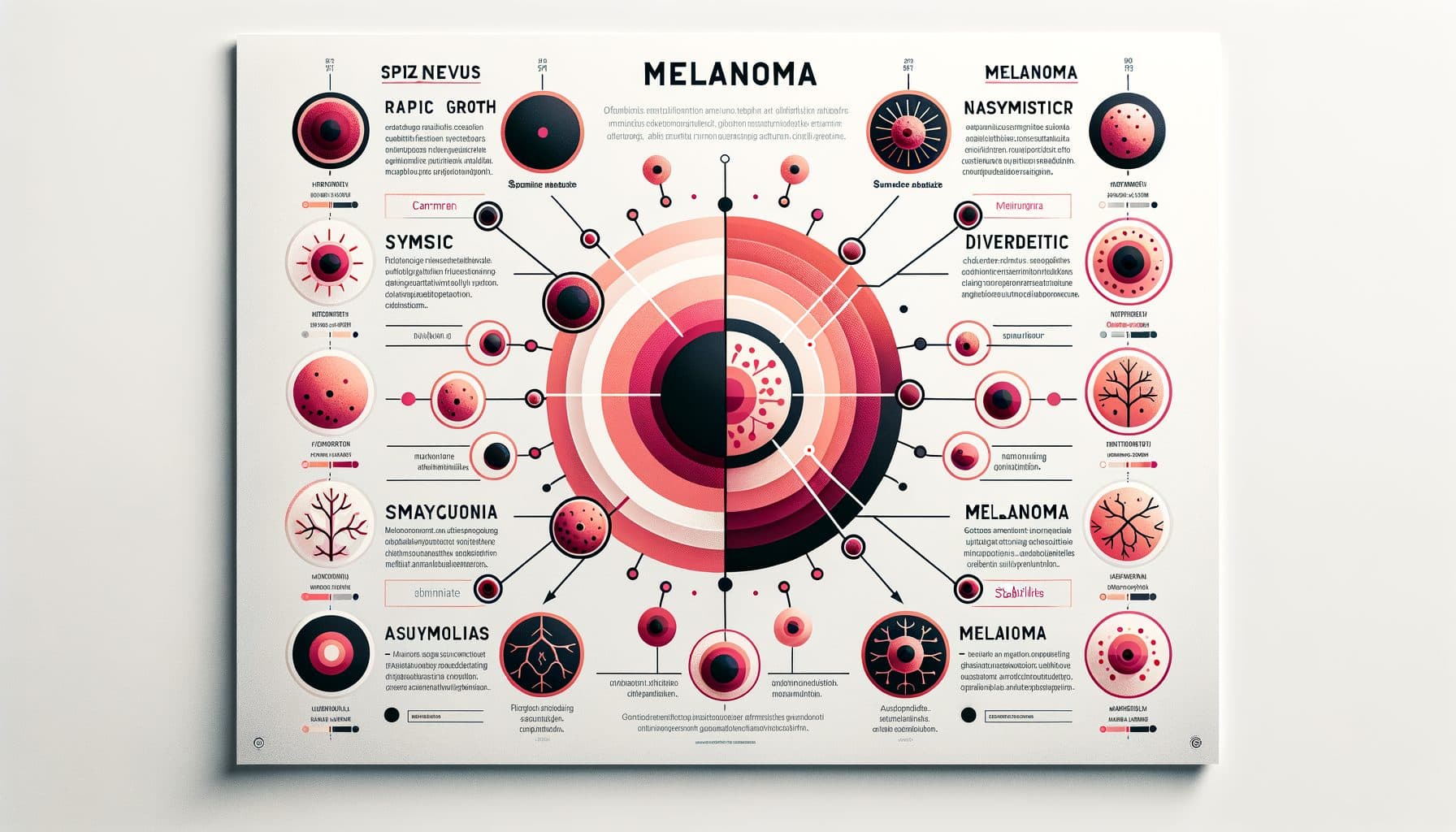
The histologic resemblance between Spitz nevi and melanoma has been a source of diagnostic controversy for more than seven decades. Under the microscope, Spitz nevi display many of the features that pathologists associate with malignancy: large, pleomorphic epithelioid and spindle cells with abundant cytoplasm and prominent nucleoli; architectural disorder with melanocytes arranged in large, confluent nests; pagetoid spread of melanocytes upward into the epidermis (a feature typically associated with melanoma in situ); high mitotic activity, particularly in rapidly growing lesions in young patients; and deep extension into the dermis. These features, seen individually or collectively, would raise serious concern for melanoma in any other context.
What distinguishes a classic Spitz nevus from melanoma histologically includes overall symmetry and circumscription of the lesion; maturation with depth (cells become smaller and less atypical in the deeper portions); the presence of characteristic Kamino bodies (eosinophilic globules at the dermo-epidermal junction); cleavage artifacts around melanocyte nests; and the absence of atypical deep mitoses. The problem is that these distinguishing features are subtle and exist on a spectrum — between a clearly benign classic Spitz nevus and an obvious melanoma lies a gray zone of lesions that experienced pathologists cannot classify with certainty. These diagnostically ambiguous lesions have been given various names including atypical Spitz tumor (AST), spitzoid melanocytic tumor of uncertain malignant potential (STUMP), and spitzoid melanocytic neoplasm of uncertain significance. The existence of this gray zone has profound clinical implications for patient management.
Spitz Nevi in Children vs. Adults
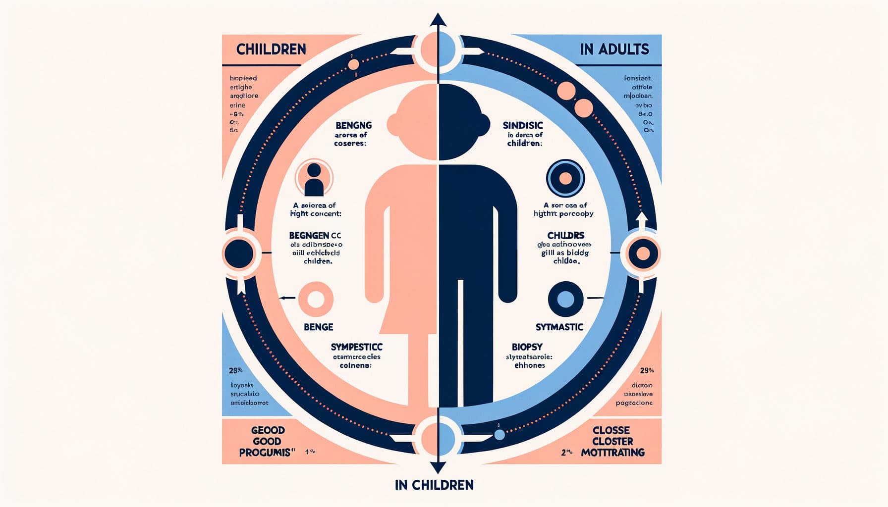
The age of the patient profoundly influences the diagnosis, prognosis, and management of Spitz nevi. In children, particularly those under the age of 12, classic Spitz nevi are common, well recognized, and overwhelmingly benign. The typical presentation — a symmetric, well-circumscribed, pink or red dome-shaped papule on the face or extremity of a young child — is clinically distinctive and pathologically reassuring.
Even when these childhood Spitz nevi show worrisome histologic features (pagetoid spread, mitoses, large size), the outcomes are almost uniformly excellent. True spitzoid melanoma in prepubertal children is extremely rare — some authorities argue it is essentially nonexistent or vanishingly uncommon.!! This favorable prognosis in children informs a more conservative management approach: many pediatric dermatologists and pathologists are comfortable with complete conservative excision and observation for classic Spitz nevi in young children.
In adults, the diagnostic landscape changes significantly. Spitz nevi become less common with increasing age while melanoma becomes more common, shifting the pretest probability toward malignancy. A spitzoid lesion in an adult — particularly over the age of 40 — is more likely to represent a spitzoid melanoma than a benign Spitz nevus.!!
Additionally, the biologic behavior of atypical spitzoid tumors in adults is less predictable than in children, with a meaningful (though still low) risk of adverse outcomes including sentinel lymph node positivity and rarely distant metastasis. Consequently, management of spitzoid lesions in adults tends to be more aggressive, with wider excision margins and more frequent use of sentinel lymph node biopsy for atypical tumors.
Diagnosis and the Atypical Spitz Tumor Controversy
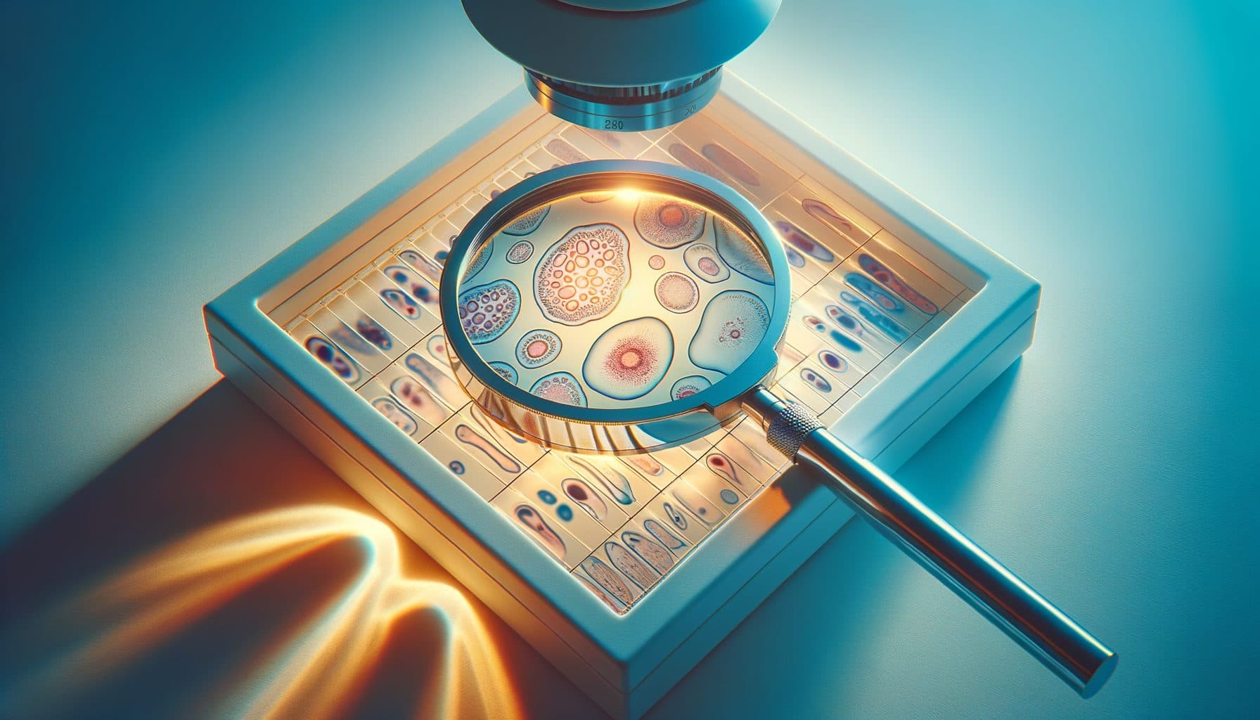
The diagnosis of Spitz nevi relies primarily on histopathological examination following biopsy, supplemented by clinical and dermoscopic assessment. Dermoscopy of classic Spitz nevi reveals characteristic patterns: the starburst pattern (radial streaming or pseudopods arranged symmetrically around the periphery) is highly specific for Spitz nevi, particularly pigmented variants. Other dermoscopic patterns include a globular pattern (symmetric brown globules), a homogeneous pattern, and an atypical pattern with multicomponent features.
The dotted vessel pattern (regularly distributed red dots) is common in non-pigmented Spitz nevi. On histopathology, an experienced dermatopathologist can usually distinguish a classic Spitz nevus from melanoma. However, the category of atypical Spitz tumor (AST) represents one of the most controversial areas in pathology.
These lesions have features that are more atypical than a classic Spitz nevus but lack sufficient criteria for a definitive diagnosis of melanoma. Different pathologists examining the same lesion may reach different conclusions — studies have shown significant interobserver variability, with some pathologists classifying a lesion as Spitz nevus while others call it melanoma. Molecular testing has improved diagnostic accuracy: fluorescence in situ hybridization (FISH) can detect chromosomal copy number changes associated with melanoma, comparative genomic hybridization (CGH) identifies broader chromosomal gains and losses, and gene expression profiling provides additional diagnostic information.
The presence of homozygous loss of 9p21 (CDKN2A), gains of 6p25 or 11q13, or complex chromosomal aberrations supports a melanoma diagnosis, while isolated kinase fusions (BRAF, ROS1, ALK, NTRK, RET, MET) without additional aberrations are characteristic of Spitz tumors. Despite these advances, definitive classification remains impossible for some lesions.
Management Approaches
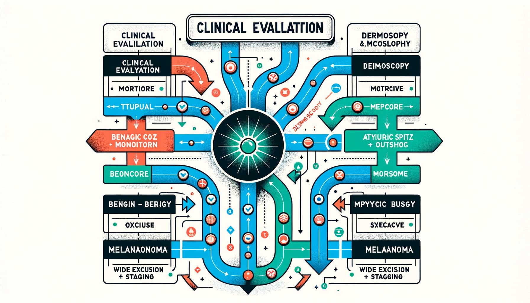
Management of Spitz nevi depends on the clinical context, age of the patient, and histopathologic classification. For a classic, small, symmetric pink papule in a child where the clinical diagnosis of Spitz nevus is confident, some experts advocate clinical monitoring without biopsy, particularly if the lesion shows a classic dermoscopic pattern. However, many clinicians prefer to biopsy any suspected Spitz nevus to obtain a definitive histologic diagnosis, especially because clinical diagnosis alone is not perfectly reliable.
Excisional biopsy — removing the entire lesion with narrow margins — is the preferred biopsy technique, as partial biopsy may not capture diagnostic features and can make histopathologic interpretation more difficult. For histologically confirmed classic Spitz nevi with reassuring features and negative margins, no further treatment is needed. Regular follow-up to monitor for recurrence is recommended, with re-excision if the lesion recurs.
For atypical Spitz tumors (ASTs), management is more debated. Current consensus for ASTs in children and adolescents favors complete re-excision with clear margins (typically five millimeters) and close clinical follow-up. Sentinel lymph node biopsy (SLNB) for ASTs in children is controversial — while up to 50 percent of ASTs may show sentinel node positivity, the clinical significance of positive nodes in pediatric atypical spitzoid tumors differs from melanoma, and most positive nodes do not lead to distant metastasis or death.
Many experts now recommend against routine SLNB for pediatric ASTs, reserving it for lesions with particularly concerning molecular or histologic features. In adults, SLNB is more commonly performed for ASTs because the risk of adverse outcomes is higher and the pretest probability of melanoma increases with age. For any lesion classified as spitzoid melanoma, management follows melanoma treatment guidelines including wide local excision with appropriate margins and sentinel lymph node biopsy.
How AI Skin Analysis Can Help
A rapidly growing, pink or pigmented nodule on a child's face or an adult's skin raises immediate concern. Skinscanner provides rapid AI-powered initial assessment when you photograph such a lesion, evaluating its visual characteristics — shape symmetry, color uniformity, border definition, surface texture, and size — against patterns associated with benign Spitz nevi and concerning features that suggest more atypical lesions or melanoma. The AI can recognize the classic presentation of a Spitz nevus — a smooth, symmetric, dome-shaped pink papule — and distinguish it from the irregular, asymmetric, multicolored appearance more suggestive of melanoma.
For pigmented variants, the app evaluates the symmetry and regularity of the pigment pattern. Skinscanner is particularly valuable as a first-line screening tool for parents who notice a new, growing lesion on their child. The app provides immediate context about what the lesion might represent, reducing anxiety when features are reassuring while appropriately recommending professional evaluation when the appearance is atypical.
For individuals monitoring existing spitzoid lesions, serial photography helps document stability or change over time — critical information for clinical decision-making. Skinscanner does not replace dermoscopic examination and histopathologic evaluation, which are essential for definitive diagnosis of spitzoid lesions. Any new, growing, or changing pigmented or pink nodule — particularly one that is asymmetric, multicolored, or larger than one centimeter — should be evaluated by a dermatologist. But Skinscanner helps bridge the gap between noticing a lesion and obtaining professional care.